|
Sample preparation and storage
Core Facility for Mass Spectrometry & Proteomics offers user-friendly protocols and provides
guidelines relevant for successful sample preparation.
Protocols for regular services
- Molecular weight determination of intact proteins (protocol)

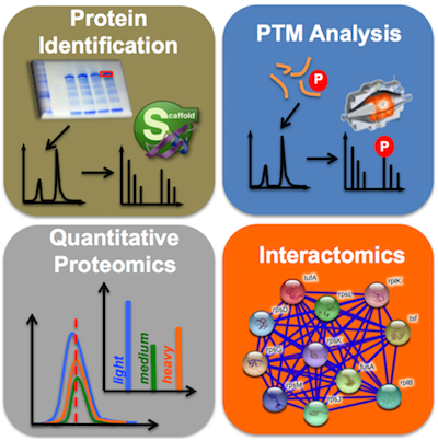
- Protein identification of gel separated proteins (protocol)
- Identification of interaction partners from Co-IP experiments (protocol)
- Analysis of proximity labeling (BioID) experiments (protocol)
- Proteome analysis (protocol)
General guidelines and practical hints relevant for sample preparation
- Contaminants may be introduced at several steps during sample preparation.
Some will go unnoticed, others will totally obscure all proteins.
Therefore, minimizing contamination is essential.
- Keratins are omni-present and notorious contaminating proteins originating from skin, hair, dust,
clothes, chemicals, etc. Total elimination is virtually impossible (and not necessary), but over-abundance will result in the repeated identification of keratins,
and not your protein of interest. Most contaminating keratins originate from dust collected during sample processing.
Therefore, a clean lab environment will certainly benefit the outcome of the MS experiment. To reduce keratin contamination, use clean containers for staining gels,
cover them with a transparent plate when inspecting them and bind your hair.
- Other frequently observed proteins are BSA, immunoglobulins, and other serum proteins. These usually originate from cell culture media,
and can be reduced by extensive washing of cell pellets with PBS prior the lysis/ protein extraction.
- In a way, polymers are more serious contaminants than keratins, since they tend to stick to HPLC columns either ruining them,
or at least causing a severe memory effect in subsequent LC-MS runs. On top of this, most polymers ionize more easily
than peptides ─ so that even minute amounts can be deleterious for the experiment. The most frequently observed polymers
are various forms of PEG, which are present in some plastics but also in soaps, hand-creams (wear gloves!) and detergents.
Don`t perform additional autoclaving of tubes or tips because that may release plasticizers which
interfere with the downstream analysis.
- One of the most pivotal steps in sample processing is the preparation of cell lysates or protein precipitates.
To do that in a reproducible way is a difficult task. Imagine that you are working with thousands of proteins with very different properties and a proteomics analysis aims to accurately quantify all of them.
- Variability in protein extraction is a major cause of low-quality proteomics data. Always try to use protocols
which are as simple as possible to increase reproducibility. We recommend to optimize your sample preparation before
preparing your sample for Proteomics.
- Sample quality, protein amount and protein extraction reproducibility can be checked using colorimetric assays for protein concentration determination, SDS PAGE/Coomassie staining and Western blotting.
If suitable antibodies are available, western blotting can be used to confirm presence of a protein of interest (e.g. bait protein) in the samples before submitting them to the facility.
Guidelines relevant for sample storage
- SDS-PAGE gels can be stored almost indefinitely in a clean plastic container, sealed bag, or wrapped
in plastic foil with a small amount of water to maintain moisture. Typically, we store our gels at 4°C.
- For cell lysates and protein extracts, it is advisable to flash freeze them in liquid nitrogen and subsequently store
them at -80°C. It is important to note that certain chemicals, such as Urea or SDS, may precipitate at lower temperatures.
Therefore, when thawing the samples, ensure that all chemicals are fully dissolved and that all proteins are properly resuspended
to prevent errors in protein concentration determination.
|