Stefan Pfeffer
ZMBH Junior Research Group Leader
ZMBH
Im Neuenheimer Feld 346
69120 Heidelberg, Germany
Tel. +49 (0) 6221 - 54 6893
s.pfeffer@zmbh.uni-heidelberg.de
|
Welcome to the Pfeffer lab!
Co-translational folding and maturation of proteins require an intricate network of folding chaperones and processing enzymes that act on the growing nascent protein in a co-translational manner and co-purify with polysomal complexes. Structural information on ribosome-nascent chain-chaperone complexes is sparse, because the involved interactions are mostly transient, labile and possibly highly flexible for chaperones binding exclusively to the growing nascent protein. This renders the involved assemblies inaccessible to classical reductionist structural biology approaches that rely on extensive biochemical purification and require conformationally homogenous particle populations for averaging. We consequently pursue a different approach and image these processes using cryo electron tomography (cryo-ET)-based strategies, which can reveal the three-dimensional arrangement of individual macromolecules even in crowded native microenvironments at molecular resolution and therefore render extensive biochemical purification unnecessary. This approach allows us to analyze the three-dimensional spatial distribution of ribosomes, chaperones and processing enzymes for individual native polysomal assemblies under conditions that preserve the labile and transient interactions governing co-translational protein folding and maturation. We study defined polysomal assemblies engaged in the synthesis of model substrates in various organisms, visualizing these assemblies in both a non-cellular context and in sections of vitrified unperturbed cells obtained using focused ion beam (FIB) milling.
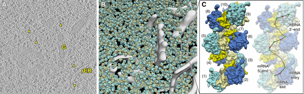
Structural analysis of polysomal assemblies in the native cellular context. (A) Slice through a representative cryo electron tomogram of an unperturbed vitrified yeast cell thinned to electron-transparent thickness using Focused Ion Beam (FIB) milling. The rough endoplasmic reticulum (rER), the Golgi apparatus (G) and several example ribosomes (arrow heads) are indicated. (B) Three-dimensional visualization of the same tomogram with cellular membranes (grey) and detected ribosomes (large subunit: cyan, small subunit: yellow) shown. (C) Example polysome within the tomogram with consecutive ribosomes numbered and depicted in alternating color. The trace of the messenger RNA (mRNA) molecule was inferred based on the positions of mRNA entry (green) and exit (red) sites on the small ribosomal subunitsext. In order to gain the most detailed structural insights possible into co-translational protein folding and maturation, we also innovate cryo-ET based data acquisition strategies and image processing schemes, optimizing them for high resolution. In particular, we integrate complementary information from different cryo-EM approaches to gain information on the three-dimensional architecture of the specimen while retaining high-resolution signal for structure determination.
Selected publications Original Research Papers Zupa, E.*, Würtz, M.*, Neuner, A., Hoffmann, T., Rettel, M., Böhler, A., Vermeulen, B.J.A., Eustermann, S#., Schiebel, E.#, Pfeffer, S.#, 2022. The augmin complex architecture reveals structural insights into microtubule branching. Nat Commun 13, 5635. Würtz, M.*, Zupa, E.*, Atorino, E.S.*, Neuner, A., Böhler, A., Rahadian, A.S., Vermeulen, B.J.A., Tonon, G., Eustermann, S., Schiebel, E.#, Pfeffer, S.#, 2022. Modular assembly of the principal microtubule nucleator gamma-TuRC. Nat Commun 13, 473. Cerullo, F.*, Filbeck S.*, Patil, P.R., Hung, H.C., Xu, H., Vornberger, J., Hofer, F.W., Schmitt, J., Kramer, G., Bukau, B., Hofmann, K., Pfeffer, S.#, Joazeiro, C.A.P#, 2022. Bacterial ribosome collision sensing by a MutS DNA repair ATPase paralogue. Nature 603, 509-14. Filbeck, S.*, Cerullo, F.*, Paternoga, H., Tsaprailis, G., Joazeiro, C.A.P.°, Pfeffer, S.°, 2020. Mimicry of canonical translation elongation underlies alanine tail synthesis in RQC. Mol Cell, in press, https://doi.org/10.1016/j.molcel.2020.11.001. Zupa, E.*, Zheng, A.*, Neuner, A., Würtz, M., Liu, P., Böhler, A., Schiebel, E.°, Pfeffer, S°, 2020. The cryo-EM structure of a gamma-TuSC elucidates architecture and regulation of minimal microtubule nucleation systems. Nat Commun 11, 5705. Wild, K., Aleksic, M., Lapouge, K., Juaire, K., Flemming, D., Pfeffer, S°, Sinning, I.°, 2020. MetAP-like Ebp1 occupies the human ribosomal tunnel exit and recruits flexible rRNA expansion segments. Nat Commun 11, 776. Liu, P.*, Zupa, E.*, Neuner, A., Böhler, A., Loerke, J., Flemming, D., Ruppert, T., Rudack, T., Peter, C., Spahn, C., Gruss, O.J., Pfeffer, S.°, Schiebel, E.°, 2019. Insights into the assembly and activation of the microtubule nucleator γ-TuRC. Nature, doi: 10.1038/s41586-019-1896-6. Martinez-Sanchez, A., Kochovski, Z., Laugks, U., Meyer Zum Alten Borgloh, J., Chakraborty, S., Pfeffer, S., Baumeister, W., Lu?i?, V., 2020. Template-free detection and classification of membrane-bound complexes in cryo-electron tomograms. Nat Methods, doi: 10.1038/s41592-019-0675-5. Schaffer, M., Pfeffer, S.*, Mahamid, J.*, Kleindiek, S.*, Laugks, T., Albert, S., Engel, B.D., Rummel, A., Smith, A.J., Baumeister, W., Plitzko, J.M., 2019. A cryo-FIB lift-out technique enables molecular-resolution cryo-ET within native Caenorhabditis elegans tissue. Nat Methods 16, 757-762. Delarue, M.*, Brittingham, G.P.*, Pfeffer, S.*, Surovtsev, I.V. , Pinglay, S., Kennedy, K.J., Schaffer, M., Gutierrez, J.I., Sang, D., Poterewicz, G., Chung, J.K., Plitzko, J.M., Groves, J.T., Jacobs-Wagner, C., Engel, B.D., Holt, L.J., 2018. mTORC1 controls phase separation and the biophysical properties of the cytoplasm by tuning crowding. Cell 174, 338-349. Braunger, K.*, Pfeffer, S.*°, Shrimal, S., Gilmore, R., Berninghausen, O., Mandon, E.C., Becker, T., Förster, F.°, Beckmann, R.°, 2018. Structural basis for coupling protein transport and N-glycosylation at the mammalian endoplasmic reticulum. Science 360, 215-219. Pfeffer, S., Dudek, J., Schaffer, M., Ng, B., Albert, S., Plitzko, J.M., Baumeister, W., Zimmermann, R., Freeze, H.H., Engel, B.D., Förster, F., 2017. Dissecting the molecular organization of the translocon-associated protein complex. Nat Commun 8, 14516. Khoshouei, M.*, Pfeffer, S.*, Baumeister, W., Förster, F., Danev, R., 2016. Subtomogram analysis using the Volta phase plate. J. Struct. Biol. 197, 94-101. Mahamid, J., Pfeffer, S., Schaffer, M., Villa, E., Danev, R., Cuellar, L.K., Förster, F., Hyman, A.A., Plitzko, J.M., Baumeister, W., 2016. Visualizing the molecular sociology at the HeLa cell nuclear periphery. Science 351, 969-972. Pfeffer, S., Burbaum, L., Unverdorben, P., Pech, M., Chen, Y., Zimmermann, R., Beckmann, R., Förster, F., 2015. Structure of the native Sec61 protein-conducting channel. Nat Commun 6, 8403. Pfeffer, S.*, Woellhaf, M.W.*, Herrmann, J.M., Förster, F., 2015. Organization of the mitochondrial translation machinery studied in situ by cryoelectron tomography. Nat Commun 6, 6019. Pfeffer, S.*, Dudek, J.*, Gogala, M., Schorr, S., Linxweiler, J., Lang, S., Becker, T., Beckmann, R., Zimmermann, R., Förster, F., 2014. Structure of the mammalian oligosaccharyl-transferase complex in the native ER protein translocon. Nat Commun 5, 3072. Pfeffer, S., Brandt, F., Hrabe, T., Lang, S., Eibauer, M., Zimmermann, R., Förster, F., 2012. Structure and 3D Arrangement of Endoplasmic Reticulum Membrane-Associated Ribosomes. Structure 20, 1508-1518. Review Articles Filbeck, S.*, Cerullo, F.*, Pfeffer, S.#, Joazeiro, C.A.P.#, 2022. Ribosome-associated quality-control mechanisms from bacteria to humans. Mol Cell 82: 1451-66 Zupa, E., Liu, P., Würtz, M., Schiebel, E.°, Pfeffer, S.°, 2020. The structure of the gamma-TuRC: a 25-years-old molecular puzzle. Curr Opin Struct Biol 66, 15-21. Pfeffer, S., Mahamid, J., 2018. Unravelling molecular complexity in structural cell biology. Curr Opin Struct Biol 52, 111-118.Pfeffer, S., Dudek, J., Zimmermann, R., Förster, F., 2016. Organization of the native ribosome-translocon complex at the mammalian endoplasmic reticulum membrane. Biochim Biophys Acta 10, 2122-2129. Pfeffer, S., Förster, F., 2016. Sec61: A static framework for membrane-protein insertion. Channels (Austin) 10, 167-169.
* equal contribution ° co-corresponding authors PhD students/postdocs from the Pfeffer lab |