
Winners of our annual Picture Award |
||||
|
2004
|
2005: Raggedy Gland
|
2006: A Scientist's Nightmare
|
||
 |
 |
 |
||
|
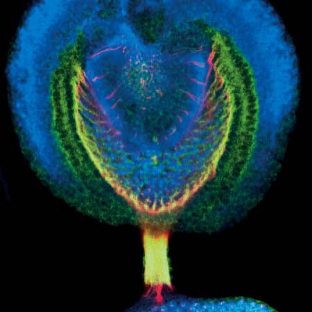
During larval development of Drosophila melanogaster, the red photo-receptors of the future retina project their axons (red) to the optic lobe to connect with neurons responsible for integration of visual information. In these axons, the protein APPL (green) is enriched. The nuclei of the neuronal cells are shown in blue. The picture was taken by Alexander Löwer (research group Paro) with the Leica Confocal Microscope TCS SP2. |
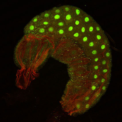
This image shows the tissue that led to the discovery of heterochromatin in Drosophila by Emil Heitz in the 1930s - a larval salivary gland. The fact that it was slightly disrupted while dissection proves that sometimes beauty does not lie in perfection. The salivary gland was stained for a wingless reporter (green) which localizes to the nuclei of secretory cells of the gland. The membrane staining is shown in red. This picture shows an optical section taken with a confocal microscope at 20x magnification by Nara Lee (research group Paro). |
by Frieder Merz (AG Bukau) ... just imaging you have a new idea, you carefully design and perform your experiments and you work hard for several days and weeks and in the end yuo face up to this. This is a memorial to all dedicated scientists to stay curious and patient. |
||
|
2007: Being Watched
|
2008: A Family Meeting
|
2009: Surprising world of lymphatic vessels
|
||
 |
 |
 |
||
|
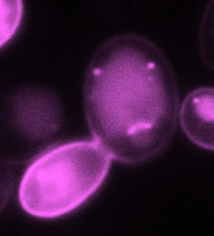
by Kiran Maaß (AG Seedorf, inverted microscope at 100x magnification) One day, after having spent too much time at the microscope the yeast cells started to be bothered by my observation... The image shows a budding yeast cell expressing the plasma membrane protein Hexose transporter 1 which is endocytosed and transported to the vacuole via endosomes. |
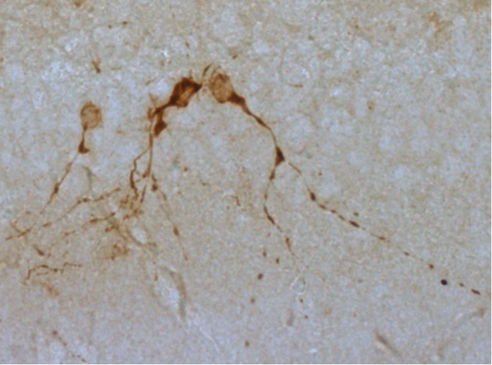
by Rosanna Parlato (AG Schütz, DKFZ-ZMBH Alliance) Newly generated nerons doublecortin positive in the dentate gyrus of a degenerating hippocampus in mutant mice lacking the transcription factor TIF-IA. Head to head fight or the peaceful encounter of a family? |
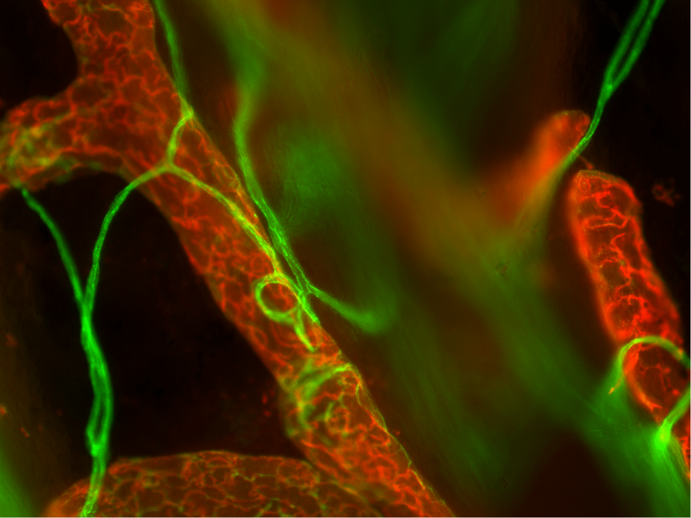
by Sila Appak (AG Augustin, DKFZ-ZMBH Alliance) 3D iimmunoflorescence picture of subcutaneous blood and lymph vessels in the mouse ear. Lymphatic vessels stained against LYVE-1 (red) are much larger than blood vessels stained against CD31 (green). The whole mount micrograph shows the expression of the endothelial cell marker CD31 (green) in lymphatic endothelial cells (red). The image was taken on an Olympus IX81 inverted microscope. |
||
|
2010: Stairway to nowhere
|
||||
 |
||||
|
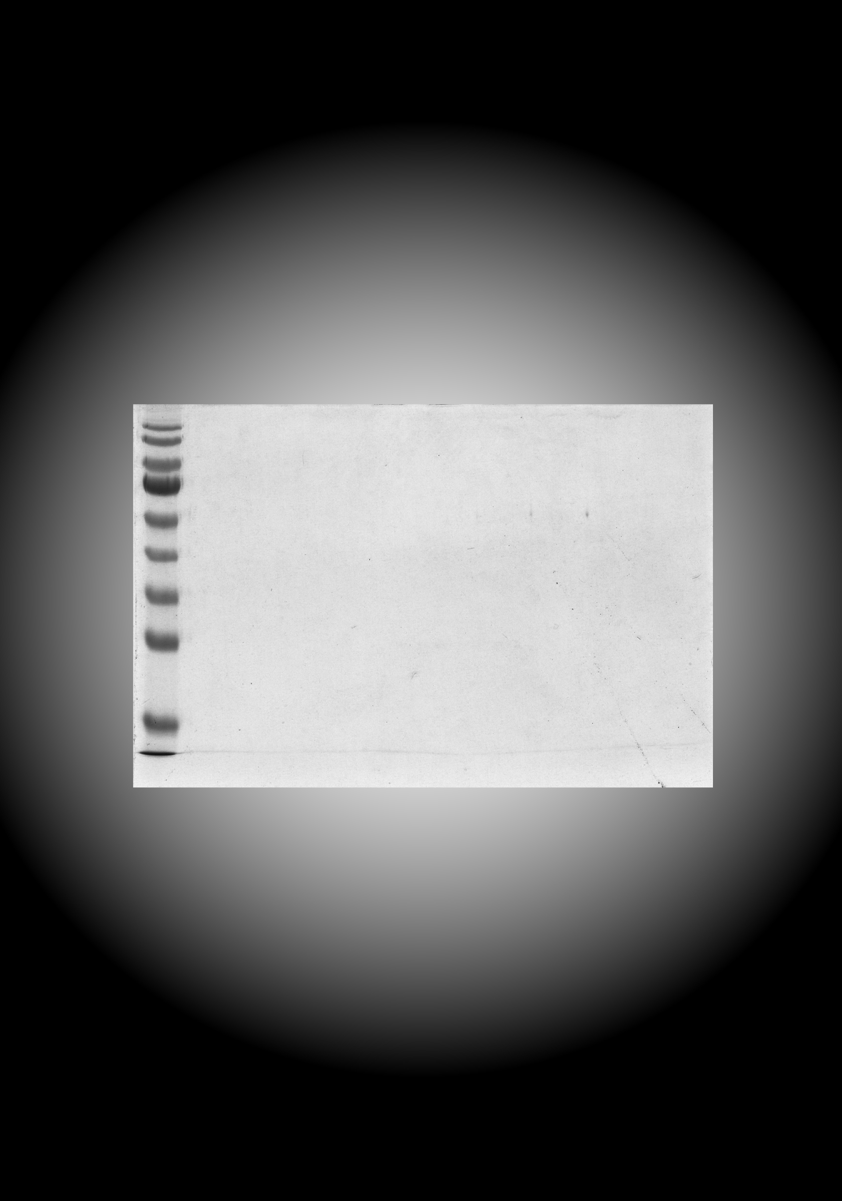
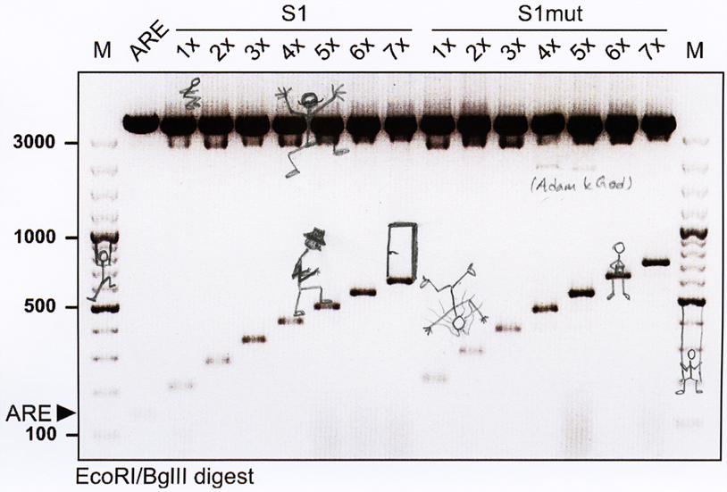
by Kathrin Leppek & Sevim Özgür (AG Stoecklin, DKFZ-ZMBH Alliance) One experiment can take the hard-working scientist to the next level. Even if it throws you back to where you started, there are always options to reach higher. You just need to keep going! This is an EcoRI/BglII-restriction digest of plasmid DNA containing repeat elements that create a staircase of fragments when excised. For this Stoecklin lab collaboration, it only took a DNA digest plus a nice agarose gel (Kathrin Leppek) as well as a very creative labmate (Sevim Özgür). |
||||
| Copyright © by the DKFZ-ZMBH Alliance. All rights reserved. |
||||