Microscopes / Instruments of the ZMBH Imaging Facility:
| Advanced confocal, STED and fluorescence lifetime imaging: |
| Leica TCS SP8 3X STED FALCON |
Microscope stand: Leica DMi8, inverted microscope
Laser:
White light laser (WLL), 470-670 nm, pulsed, no pulse picker
STED lasers for superresolution:
592 nm, 1.5 W
660 nm, 1.5 W
775 nm, >1.5 W
Objectives:
HC PL FLUOTAR 10x/0.30 Air
HC PL APO 20x/0.75 IMM CORR Lbd BL
HCX PL APO 63x/1.40 Oil CS2
HC PL APO 100x/1.40 STED White Oil
2 Hybrid GaAsp detectors (1st gen HyDs) and 2 PMTs
gated STED
3D STED 3X
Software: Leica Application Suite X (LASX)
FALCON for FLIM, FLIM-FRET, TauSTED and Phasor
Room 122, INF 345
|
Dedicated for advanced applications: Superresolution (3D) STED, fluorescence lifetime applications: FLIM, fitting-based and Phasor-based FLIM analysis, FLIM-FRET, tauSTED, high-resolution confocal plus deconvolution.
| see instrument |
| Point-scanning confocal microscopy: |
| Leica TCS SP5 |
Microscope stand: Leica DMi8, inverted microscope
Microscope stand: Leica DMI 6000 CS Trino, inverted microscope
Laser:
405 nm diode laser, 50 mW
458 nm, 5 mW, 476 nm, 5mW, 488 nm, 20 mW, 514 nm, 20 mW Argon laser
561 nm DPSS laser, 20 mW
633 nm HeNe laser, 10 mW
Objectives:
HC PL APO 10x/0.40 CS Air
HCX PL APO 20x/0.70 IMM CORR Lbd BL
HCX Plan APO 40x/1.30 Oil CS
HCX PL APO 63x/1.30 GLYC CORR CS
HCX PL APO 63x/1.40-0.60 Oil Lbd BL
2 Hybrid GaAsp detectors (1st gen HyDs) and 3 PMTs
Tandem scanning system
Incubation chamber w/ Temp & CO2 control (LIS)
Software: Leica Application Suite (LAS)
AFC (hardware autofocus)
Room 128, INF 345
Point-scanning confocal microscope for fixed and live samples. Equipped with temperature and gas control, hardware autofocus and tandem scanner. The motorized stage allows for 2D/3D tile scan and mark-and-find applications.
| see instrument |
|
| Zeiss LSM 780 |
Microscope stand: Zeiss Axio Observer, inverted microscope
Laser:
405 nm pulsed diode laser, 30 mW
458 nm, 488 nm, 514 nm Argon laser, 25 mW
561 nm DPSS laser, 20 mW
633 nm HeNe laser, 5 mW
(pulsed) in Tune laser 490-640 nm, 1.5 mW per wavelengthObjectives:
Plan-APOCHROMAT 20x/0,80 Air
C-APOCHROMAT 40x/1.2 W Korr-UV-VIS-IR
Plan-APOCHROMAT 40x/1.3 Oil
Plan-APOCHROMAT 63x/1.4 Oil
Plan-APOCHROMAT 100x/1.4 Oil
Software: Zeiss ZEN 2010
Room 151, INF 329
Point-scanning confocal microscope for fixed samples and short-term live observations. The manual microscope stage is easily accessible, i.e., an 'open system' without an incubation chamber. The inTune-laser enables the precise excitation of non-common dyes/labels with individual wavelength settings (small step sizes) between 490nm and 640nm. Besides standard confocal applications (includung FRAP, FLIP), also F(C)CS and RICS can be performed at the system.
| see instrument |
| Spinning disk confocal microscopy: |
| Leica DMi8 Spinning Disk microscope |
Microscope stand: Leica DMi8, inverted microscope
Confocal Scanner Unit: Yokogawa CSU-X1
Laser:
405 nm, diode laser
488 nm, 50 mW diode laser
561 nm, 50 mW diode laser
640 nm, diode laser
Objectives:
HC PL APO 20x/0.7 Air
HC PL FLUOTAR 25x/0.8 IMM CORR
HCX PL S-APO 40x/0.75 Air
HC PL APO 40x/1.3 Oil
HC PL APO 63x/1.40-0.60 Oil
HC PL APO 100x/1.4 Oil CS2
Cameras:
Hamamatsu EMCCD ImagEM Enhanced (C9100-13)
Hamamatsu Orca Flash 4.0 LT (C11440-42U)
Adaptive Focus Control (AFC)
Software: MetaMorph Advanced Acquisition
Incubation chamber XL & Pecon temperature control unit
Room 155, INF 329
Best suited for fast and gentle, i.e. low phototoxicity, live cell confocal observations. The software plus the motorized stage, hardware autofocus and incubation chamber enables 3D confocal imaging of up to 4 channels over time at different locations. A dedicated module for deconvolution (SVI Huygens Professional) of the spinning disk data is available.
| see instrument |
|
| Total internal reflection fluorescence (TIRF), localization microscopy: |
| Olympus cellSens TIRF microscope |
Microscope stand: Olympus IX81, inverted microscope
Olympus cellTIRF 4-Line Motorized TIRF Combiner
TIRF Lasers:
405 nm, 100 mW
488 nm, 100 mW
561 nm, 100 mW
640 nm, 140 mWOlympus cellFRAP module
Objectives:
UPLSAPO 60x/1.35 Oil DIC
PLAPO 100x/1.45 Oil DIC
Zeiss EC Plan NeoFluar 40x/1.3 Oil
Lumencor Spectra X light engine
Filters:
Dualband CFP/YFP sbx HC filter set
Dualband GFP/mCherry sbx ET filter set
Quad DAPI/FITC/Cy3(Texas Red)/Cy5 sbx HC filter set
TIRF Quad
Cameras:
Olympus XM10
Hamamatsu ORCA-Flash4.0 (C11440)
(optional)Hamamatsu ImagEM Enhanced (C9100-13)
Software: Olympus cellSens
ZDC (hardware autofocus)
(optional) Incubation chamber w/ Temp and CO2 control units
TMC Linos optical table 63-550
Room 158, INF 329
Fully automatable TIRF microscope for up to 4 individually adjustable TIRF lines. The microscope is equipped for 3D localization microscopy (PALM, (d)STORM,..) by using a bespoke astigmatic lens.
| see instrument |
|
| Screening microscopy: |
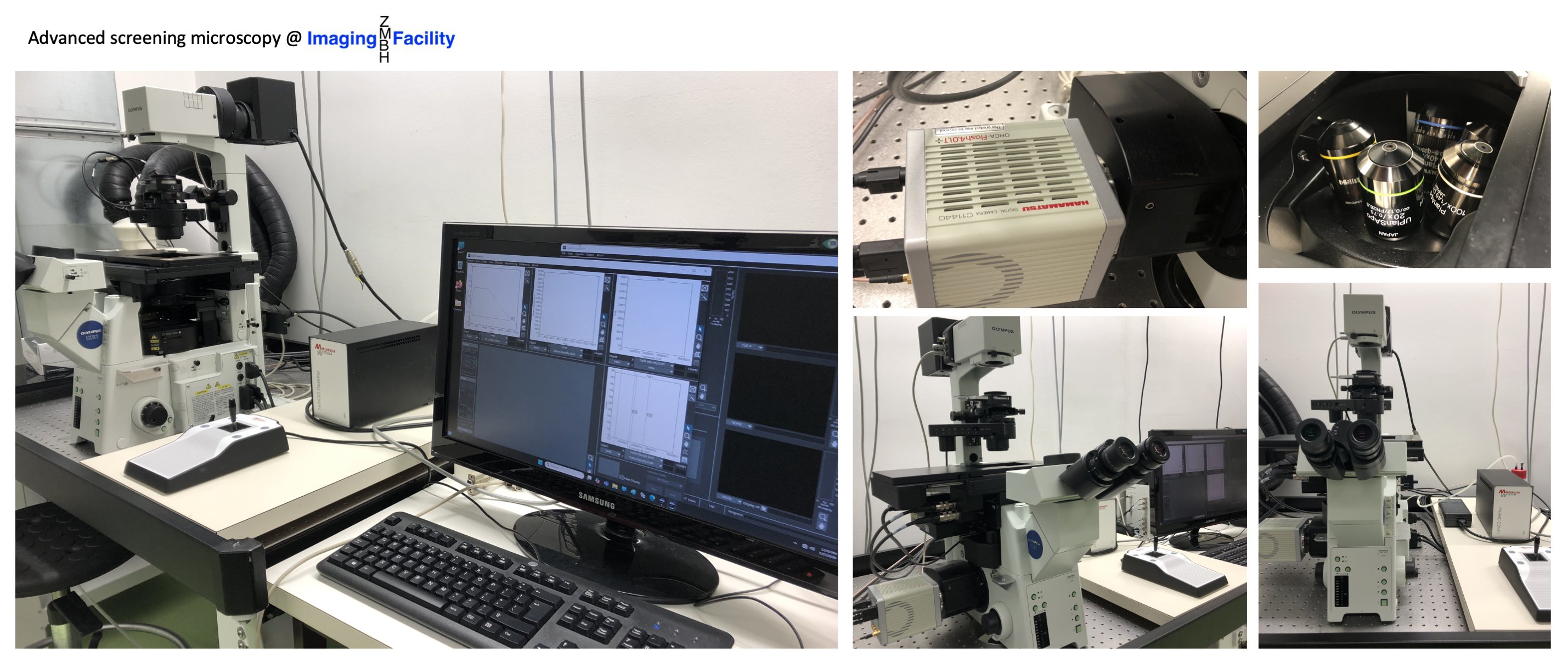
| Olympus ScanR microscope |
Microscope stand: Olympus IX81, inverted microscope
Objectives:
UPLSAPO 10x/0.40 Air
UPLSAPO 20x/0.75 Air
UPLSAPO 40x/0.95 Air
UPLFL N 60x/0.90 Air
PLAPO 100x/1.45 Oil
Lumencor Spectra X light engine
Filters for:
DAPI
CFP
GFP
YFP
RFP/mCherry
Cy5
Camera: Hamamatsu Orca Flash 4.0
Software: Olympus ScanR_2.5.0 & CellSens
ZDC (hardware autofocus)
(optional) Incubation chamber w/ Pecon temp control
Room 158, INF 329
Screening microscope for automated 2D/3D multi-well plate imaging (up to 384 well plates) including sophisticated software for data analysis. Workflows for deconvolution of the image data (batch processing) have been established by the ZMBH Imaging Facility team.
| see instrument |
|
| Live-cell imaging and analysis: |
| Incucyte S3 |
Complete S3 system located in incubator, remotely controlled
2 color green, red fluorescence, and phase contrast
Objectives:
4X, 10x, 20x
3 vessel trays and numerous vessels
Incubator for mammalian (and insect) cells
Room 103, INF 329
For long-term fully automated 2D live-cell imaging in numerous multi-well plates and dish formats. Scan Types: Standard, Image Lock, Scratch Wound, Whole Well, Dilution Cloning and Spheroid. Image analysis with the Basic Analyzer. The scratch wound device for efficient scratch-making in 96 well plates is available upon request.
| see instrument |
|
| Nikon BioStation IM-Q |
For glass bottom dishes
Objectives:
40x (20x, 40x, 80x by switching tube lens), NA 0.8,
Filters:
Ex 438/24, Em 483/32 CFP/Cerulean
Ex 472/30, Em 520/35 GFP
Ex 500/24, Em 542/27 YFP
Ex 562/40, Em 624/40 Alexa568/mCherry/TxRed
Camera: 1.3-megapixel cooled monochrome camera
Software: Nikon
Room 151, INF 329
For long-term fully automated 2D and 3D fluoresence and brightfield live-cell imaging in 35 mm-dishes and chambered coverslips.
| see instrument |
|
| Advanced widefield microscopy: |
| Olympus cellSens microscope |
Microscope stand: Olympus IX81, inverted microscope
Objectives:
UPlanSApo 4X/0.16 Air
UPLSAPO 10x/0.4 Air
UPLSAPO 20x/0.75 Air
UPLSAPO 40x/0.95 Air DIC
UPLSAPO 60x/1.35 Oil
PLAPO 100x/1.45 Oil DIC
Lumencor Spectra X illumination system
Filters:
Dualband CFP/YFP sbx HC filter set
Dualband GFP/mCherry sbx ET filter set
Quad DAPI/FITC/Cy3(Texas Red)/Cy5 sbx HC filter set
Single band GFP
Single band mCherry
Camera: Hamamatsu ORCA-R2
Software: Olympus cellSens
ZDC (hardware autofocus)
Room 123, INF 329
Versatile fluorescence microscope for a broad range of widefield applications in 2D and 3D including tile scanning. Dedicated deconvolution software (SVI Huygens Professional) is available upon request.
| see instrument |
|
| Basic widefield microscopy: |
| Leica DM5000B microscope |
Microscope stand: DM5000B, upright microscope
Objectives:
HCX PlApo 40x/1.25-0.75 Oil
HCX PlApo 63x/1.40-0.60 Oil
Camera: Andor DR-328G-CO2-SIL
Software: Leica Application Suite (LAS)
Room 158, INF 329
Basic fluorescence widefield applications, practical course microscope.
| see instrument |
|
| Olympus BX60 |
Microscope stand: Olympus BX60, upright microscope
Objectives:
Plan 4x/0.10 Air
Plan NeoFluar 10x/0.3 Air
UPlan Fl N 20x/0.5 Air
UPlanApo 40x/1.00 Oil
PlanApo 60x/1.40 Oil
UPlanSApo 100x/1.40 Oil
Camera: ProgRes MF
Software: ImageJ ProgRes camera control
Room 158, INF 329
Basic fluorescence widefield applications, practical course microscope.
| see instrument |
|
| Olympus IX71 |
Microscope stand: IX71, inverted microscope
Objectives:
UPlanFl 10x/0.30 Air Ph1
LC PlanFl 20x/0.40 Air Ph1
LC PlanFl 40x/0.60 Air Ph2
PUPlanSApo 100x/1.40 Oi
Software: Olympus Sob Imaging Systems CellF 2.8
Room 158, INF 329
Basic fluorescence widefield applications, practical course microscope.
| see instrument |
|
| Fluorescence stereo microscopy: |
| Leica M205 FA microscope |
Fully motorized fluorescence stereo microscope
Objectives:
Plan Apo 0.63x
Plan Apo 1.60x
Camera: Leica DFC300FX
Software: Leica Application Suite (LAS) Core
Room 155, INF 329
Fully motorized acquisition with color camera, for fluorescence and brightfield stereo microscopy and macroscopic samples
| see instrument |
|
| Leica M420 |
Cold light sources: Leica KL 1500 LCD & Zeiss KL 2500 LCD
Camera: ProgRes C10plus (optional)
Software: Micro-Manager (optional)
Room 155, INF 329
For macroscopic samples (flies, insects..).
| see instrument |
|
| Micromanipulation/-injection microscopy: |
| AIS 2 micromanipulation system |
Microscope stand: Zeiss Axiovert 10, inverted microscope
Injection system: Luigs & Neumann, Germany
Injection pressure system: Eppendorf FemtoJet
Objectives:
ACHROSTIGMAT 5x/0.12 Air
ACHROSTIGMAT 10x/0.25 Air
ACHROSTIGMAT 20x/0.30 Air Ph1
ACHROSTIGMAT 32x/0.40 Air Ph1
Plan NEOFLUAR 40x/0.75 AirFilters:
F41-017 HQ-Set EGFP
F41-021 HQ-Set Cy 3,5
Camera: ImagingSource DMK2002
Software: AIS 2 control program
Micromanipulation sytsem for injections of volumes into cells and removal of cytosol/cellular components from cells.
| see instrument |
|
|
|